

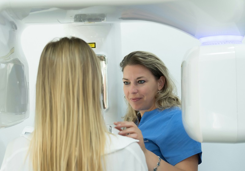
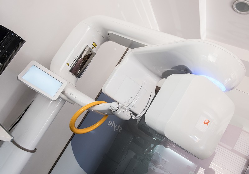
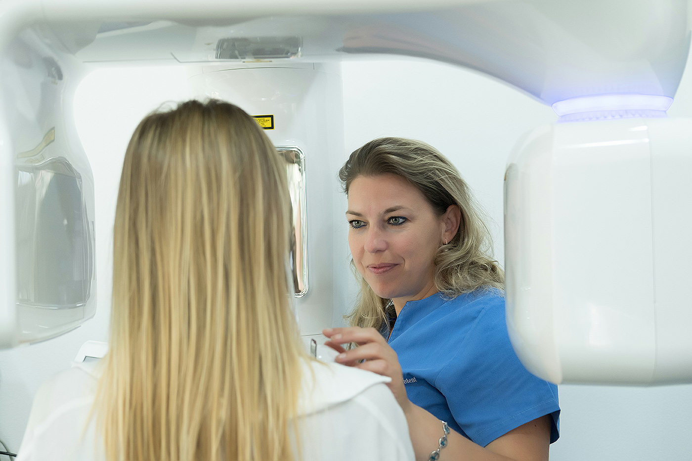
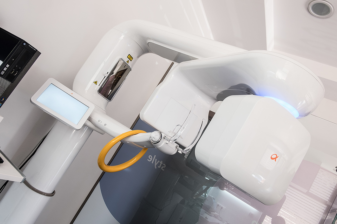
X-RAY orthopanomogram
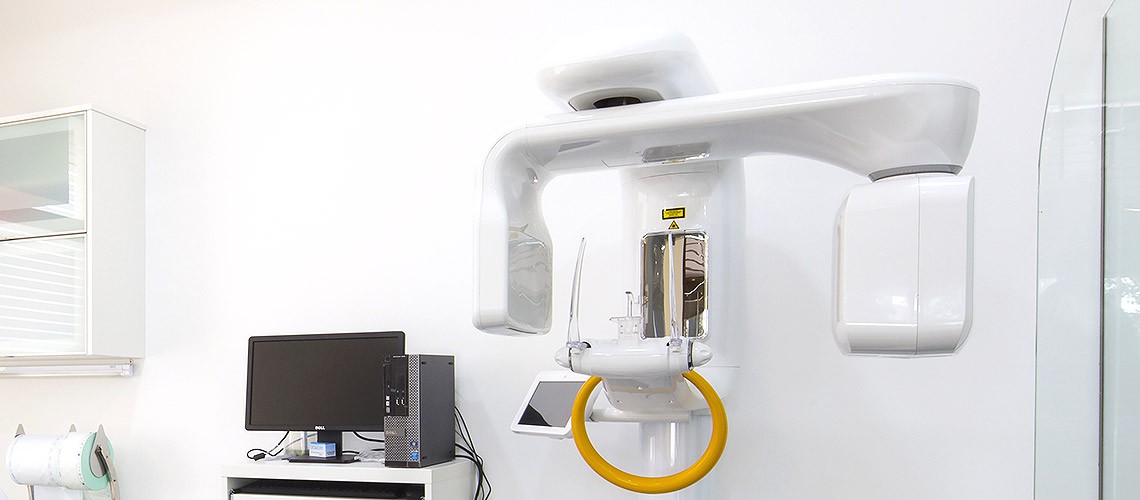
X-ray images of teeth and jaw are the most common diagnostic methods enabling the doctors of dental medicine to have an insight into the condition of structures that are not visible by clinical examination in the office. Today's digital technology enables us very precise and high-quality images with very low, almost negligible radiation dose. The technology we use at our clinic guarantees a minimum exposure to ionizing radiation for necessary examinations. The use of super sensitive sensors and special algorithms results in a significant reduction of the radiation dose necessary for imaging. Only digital technology is applied at our clinic.
The amount of radiation necessary for orthopan imaging of an average adult person is equivalent to the amount of everyday radiation we receive during one day. The amount of radiation is adjusted for each patient individually, in order to receive the lowest possible dose. Special imaging programs intended for children and their bone composition enable us the minimum dose of radiation for our youngest patients.
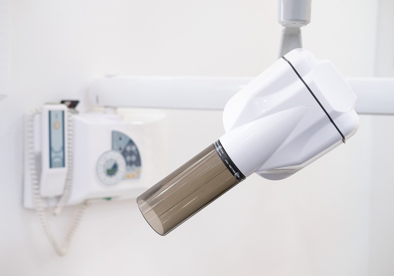
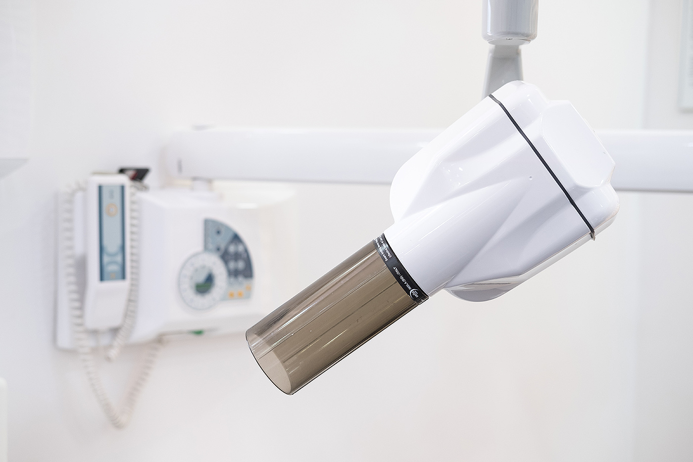
Retroalveolar images are obtained by the use of sensor placed in a patient's mouth. The position and size of sensor depend on indication.
Retroalveolar image gives us an insight into condition of 1-3 teeth. Due to its minimal radiation, it is often used in dentistry in diagnostics for various tooth disorders and diseases.
Bitewing image
Bitewing image is used for in caries diagnosis, in spaces between two teeth. The image shows crowns of upper and lower teeth of the target area.
At our clinic, we utilize an Intra Oral X-Ray Unit by Cefla and the latest digital orthopantomogram innovation RayScan by Samsung.
Book your appointment now!
+382 067 211 777 Contact us
Our partners


Copyright © 2026. Dental Montenegro. Cookies Use Policy. Privacy Policy.
All Rights Reserved. Designed by Minmedia.